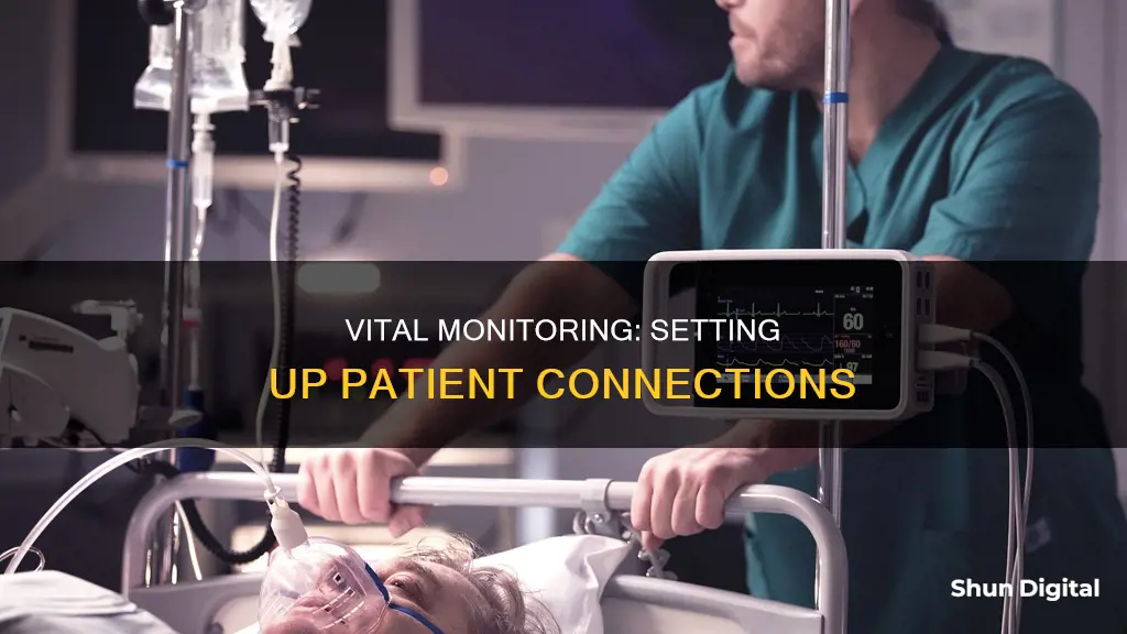
Monitoring patients is a crucial aspect of healthcare, especially in intensive care units, progressive care units, and emergency departments. Continuous cardiac electrophysiological monitoring is routinely performed for acute and critically ill patients. This involves the use of electrocardiograms (ECGs) to record electrical activity in the heart, helping to identify issues such as arrhythmias, palpitations, and cardiac problems. The placement of electrodes on specific areas of the chest is key to obtaining accurate data, and different lead systems, such as three-lead, five-lead, and six-lead configurations, are used depending on the situation. Proper placement of electrodes is essential to ensure accurate interpretation of the ECG waveform and to avoid misdiagnosis or inappropriate treatment.
| Characteristics | Values |
|---|---|
| Purpose | To monitor the electrical activity of the heart over 24 hours or longer while the patient is away from the healthcare provider's office |
| Type of Monitor | Portable electrocardiogram (ECG) |
| Components | Small, plastic patches (electrodes), wires, and a monitor box (portable recording device) |
| Electrode Placement | Chest and belly (abdomen) |
| Monitoring Duration | 24 to 48 hours, sometimes longer |
| Diary | Patients are instructed to keep a diary of their activities and symptoms during the test |
| Personal Care Instructions | Keep the device dry while it is being worn |
| Battery | May need to be changed during the monitoring period |
What You'll Learn

Prepare the patient's skin
The first step in preparing the patient's skin for a Holter monitor is to wash the skin with soap and water to ensure that the adhesive pads will stick properly. It is important to dry the skin thoroughly after washing, as moist skin can hinder electrode adherence. If the patient has thick chest hair, it may be necessary to shave the areas where the electrodes will be placed to ensure a clear reading.
Once the skin is clean and dry, the next step is to identify the sternal angle or angle of Louis. This can be done by palpating the upper sternum to locate the juncture of the clavicle and the sternum, known as the suprasternal notch. From there, slide your fingers down the center of the sternum to locate the obvious bony prominence, which is the sternal angle. This identifies the second rib and provides a landmark for locating the fourth intercostal space (ICS) for accurate electrode placement.
Next, use skin abrader or surgical clippers to clip or shave chest hair as needed to ensure good skin contact with the electrodes. It is important to remove any obstacles that may interfere with the transmission of electrical impulses.
Now, test the electrode pads for moisture by removing the backing from the pre-gelled electrodes. It is important to ensure that the gel is moist to allow for proper impulse transmission. If the gel has dried out, the electrodes may not adhere properly and could interfere with the transmission of electrical impulses.
After confirming the moisture of the gel pads, it is time to place the electrodes on the patient's skin. Apply the electrodes by pressing around their entire edges, avoiding direct pressure on the gel pads. This step is crucial as it ensures that the electrodes are securely attached and prevents external influences from affecting the ECG readings.
For a three-lead system, apply the RA electrode below the clavicle, close to the patient's right shoulder. The LA electrode should be placed near the left shoulder, and the LL electrode below the level of the umbilicus on the left abdomen.
For a five-lead system, apply the RA and LA electrodes close to the right and left shoulders, respectively, below the clavicle. The RL electrode should be placed below the rib cage on the right side, and the LL electrode on the left side. The chest lead electrode can be placed on either the V1 or V6 position, depending on the patient's needs.
By following these steps, you can effectively prepare a patient's skin for monitoring, ensuring accurate and reliable readings.
ASUS LCD Monitor: Pixelated Display Issues Explained
You may want to see also

Attach the electrodes
Attaching the Electrodes
The first step in attaching the Holter monitor is to prepare the patient's skin. Wash the chest and belly (abdomen) area with soap and water, and dry it thoroughly with gauze pads or a washcloth. This ensures that the electrodes will stick properly to the skin. Do not use alcohol for skin preparation as it can dry out the skin. If the patient has thick chest hair, clip or shave the areas where the electrodes will be placed to ensure a clear reading.
Next, open the packaging of the pre-gelled electrodes and test the centre of the pads for moisture. It is important to use these electrodes soon after opening as extended exposure to air can dry them out, reducing their adhesive and conductive properties.
Now you are ready to attach the electrodes to the patient's skin. The exact placement will depend on the type of monitor being used, but a typical configuration involves placing two electrodes on either side of the heart, one in front of the heart, and two pads on the bottom edge of either side of the rib cage. Press around the entire edges of the electrodes to ensure a tight seal, being careful not to press directly on the gel pads as this may cause the gel to leak.
Once the electrodes are securely in place, connect the leads (wires) to the electrodes according to the instructions provided. Typically, the leads snap into place on the adhesive pads. Ensure that the wires are colour-coded and plugged into the correct openings on the monitor. For example, the right arm lead, marked RA, is usually white, while the left arm lead, marked LA, is usually black.
After all the electrodes and leads are connected, check the ECG tracing on the monitor to ensure a clear pattern is displayed. The R wave should be approximately twice the height of the other components of the ECG for proper detection by the heart rate counter.
It is important to remind the patient not to shower, swim or engage in excessive sweating while wearing the Holter monitor, as this can cause the electrodes to loosen or come off.
Expanding Your View: Increasing Monitor Size in Adobe SpeedGrade
You may want to see also

Activate the monitor
To activate the monitor, you must first ensure the battery is charged and the activity lights are flashing. The monitor will record continuously and automatically turn off once the prescription period is over. You will know the monitor is working when you see the activity lights flashing.
There are various types and brands of monitors, so instructions for activation may vary. Always follow the instructions provided by your doctor.
If you are using a Holter monitor, you will need to attach the electrodes to your skin. Put the adhesive electrodes on as directed—this usually means two electrodes on either side of your heart, one in front of your heart, and two pads on the bottom edge of either side of your rib cage. Secure the leads (wires) to the electrodes according to the instructions. Typically, the leads snap into place on the adhesive pads.
Setting Up Your ASUS Monitor: A Step-by-Step Guide
You may want to see also

Keep a diary of symptoms
Keeping a diary of symptoms is an important part of the Holter monitor process. This is a small, portable electrocardiogram (ECG) that records the electrical activity of the heart over 24 hours or more. It is used to help diagnose irregular cardiac symptoms, and patients will usually wear the monitor for 24 to 48 hours.
During this time, it is important to keep a diary of symptoms and activities. This will give the doctor a more complete picture of the patient's cardiac health. The diary will include the date and time of activities, and any symptoms that occur, such as chest pain, dizziness, shortness of breath, or irregular heartbeats. It is also important to note down any activities that may affect the monitor's readings, such as being near magnets, metal detectors, or electrical appliances.
The doctor may provide a diary to fill out, or the monitor may come with a separate device that allows the patient to "track" their symptoms. This will alert the doctor to pay special attention to certain times on the monitor.
After the monitoring period is over, the patient will return the monitor and the diary to the doctor, who will then interpret the results and discuss them with the patient.
Pixel Size: Monitor Viewing and Resolution Clarity
You may want to see also

Return the monitor
Returning a patient monitor is a straightforward process, but there are a few important steps to follow to ensure the equipment is handled correctly and safely. Here is a detailed guide on how to return the monitor after use:
- Remove the monitor from your body: Once the data measurement period is complete, carefully remove the monitor by gently peeling off the electrodes from your skin. Place the device back into its original box or packaging, ensuring all components are securely packed. If there is any residue or adhesive marks on your skin from the electrodes, you can use a solvent provided by your doctor or wipe the area with water, alcohol, mineral oil, or lotion to remove any remaining stickiness.
- Return the equipment: It is essential to understand that the monitor is typically rented from the doctor's office and needs to be returned promptly. Contact the cardiologist's office to determine the specified time frame for returning the equipment. You may be required to physically bring the monitor back to their office, or they may allow you to mail it back. Ensure you also return any diary or logbook containing records of your symptoms and activities during the monitoring period.
- Schedule a follow-up appointment: After returning the monitor, schedule a follow-up appointment with your cardiologist to review the results. They will interpret the data collected by the monitor and discuss any findings and potential treatment options with you. This appointment is crucial in understanding your cardiac health and addressing any concerns or issues detected during the monitoring period.
- Proper disposal of biohazardous materials: If the monitor or any of its components contain biohazardous materials, such as blood or other bodily fluids, special disposal procedures must be followed. Contact the medical facility or refer to their guidelines for instructions on how to safely dispose of or return biohazardous waste. This ensures the protection of both medical staff and the environment.
- Authorisation for shipping: In some cases, you may need to obtain authorisation from the medical facility before shipping the monitor back. Contact the patient or customer services department, specialising in your type of device, to initiate the return process. They will provide you with the necessary instructions and, depending on the device, either send you a prepaid shipping label or a box with a pre-addressed label for returning the monitor.
- Returning explanted devices: If the monitor was implanted within the body, special precautions must be taken for its return. Contact the medical device company, such as Medtronic, to request a biohazard return kit. They will provide instructions for safely shipping the device back. Ensure you use the provided biohazard container and follow their guidelines to avoid any risks associated with handling biohazardous materials.
By following these steps, you can ensure the proper return of the patient monitor, allowing for accurate diagnostics, timely equipment turnover, and adherence to safety protocols.
Ultimate Monitor Size for the Nintendo Unisystem Arcade
You may want to see also
Frequently asked questions
A Holter monitor is a type of portable electrocardiogram (ECG) that records the electrical activity of the heart over 24 hours or more while the patient is away from the healthcare provider's office. It is used to evaluate symptoms that may be heart-rhythm related, identify irregular heartbeats, and assess the risk of future heart-related events.
Before attaching the monitor, ensure the patient's skin is washed and clean so that the adhesive pads will stick to their body. Remove any objects that may interfere with the recording, such as jewellery, and ask the patient to remove clothing from the waist up.
Place the adhesive electrodes on the patient's chest and abdomen as directed. Typically, this involves placing two electrodes on either side of the heart, one in front of the heart, and two pads on the bottom edge of either side of the rib cage. Connect the wires from the electrodes to the monitor.
Patients should be instructed to continue their normal daily activities to get an accurate picture of their heart activity. They should avoid getting the monitor wet, so they should remove it before showering or engaging in sweaty exercise. Patients may also be asked to keep a diary of their activities and any cardiac symptoms they experience.


