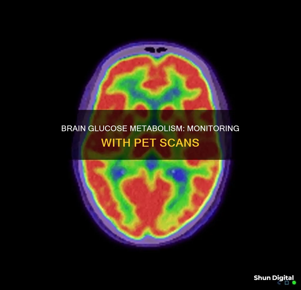
The brain uses glucose as its main source of energy. Glucose monitoring can be done through blood tests, capillary blood glucose tests, and continuous glucose monitoring.
Blood glucose tests involve taking a blood sample from a vein in the arm and are used to find out if blood sugar levels are in a healthy range.
Capillary blood glucose tests involve collecting a blood drop sample from a fingertip prick and are often referred to as capillary blood glucose (CBG) tests.
Continuous glucose monitoring (CGM) involves applying a water-resistant disposable sensor on the back of the upper arm or abdomen and can store up to 90 days of glucose data.
PET scans are also used to monitor glucose uptake in the brain.
| Characteristics | Value |
|---|---|
| --- | --- |
| Test Name | Blood Glucose Test |
| Test Type | Blood Test |
| Test Purpose | To find out if your blood sugar levels are in a healthy range |
| Test Procedure | A health care professional will take a blood sample from a vein in your arm, using a small needle. |
| Test Results | Normal range: 4 to 6 mmol/L or 72 to 108 mg/dL |
What You'll Learn
- Glucose is the brain's main source of energy
- Glucose enters the brain through facilitated diffusion via glucose transporters in the blood-brain barrier
- Brain microdialysis can be used to monitor the metabolic state of the injured brain
- Insulin therapy has been used to treat hyperglycemia in neurointensive care with conflicting results
- Monitoring of brain glucose with microdialysis has been shown to predict ischemic infarcts and detect glucose zero values despite normal blood glucose

Glucose is the brain's main source of energy
The brain's glucose supply is regulated by neurovascular coupling and may be modulated by metabolism-dependent and -independent mechanisms. Glucose enters the brain from the blood by crossing the blood-brain barrier through glucose transporter 1 (GLUT1). Glucose is then rapidly distributed through a highly coupled metabolic network of brain cells. Glucose provides the energy for neurotransmission, and several glucose-metabolising enzymes control cellular survival.
The brain's glucose usage can be monitored through a blood glucose test, which measures the glucose levels in the blood. Glucose is a type of sugar, and too much or too little glucose in the blood can be a sign of a serious medical condition. A blood sample will be taken from a vein in the arm, using a small needle.
The most common procedure that monitors glucose uptake in the brain is the PET scan.
Monitoring Data Usage on Your iPad: A Guide
You may want to see also

Glucose enters the brain through facilitated diffusion via glucose transporters in the blood-brain barrier
Glucose transporters are membrane proteins that facilitate the entry of glucose into cells, including those in the brain. Glucose transporters are encoded by twelve different genes and play a crucial role in the specific uptake of glucose by different cell types in the brain, such as endothelial cells, astrocytes, and neurons.
The glucose transporter specific to neurons is GLUT3. Its cellular distribution appears to predominate in the cell bodies rather than in the axon terminal compartment.
The glucose transporter GLUT1 is responsible for the entry of glucose into the brain. It is selectively and highly expressed on both luminal and abluminal membranes of capillary endothelial cells covering most neuronal areas.
CenturyLink Internet Usage: Monitor, Track, and Manage Your Data
You may want to see also

Brain microdialysis can be used to monitor the metabolic state of the injured brain
Brain microdialysis has been used to monitor patients with traumatic brain injury (TBI), subarachnoid hemorrhage (SAH), and ischemic stroke. In TBI and SAH patients, low cerebral glucose levels detected by microdialysis have been associated with poor neurological outcome. Monitoring brain glucose levels in these patients can provide valuable information on the development of secondary ischemia.
In addition, the location of the catheter is important for correlating brain glucose levels with plasma glucose levels. Monitoring brain glucose levels in the neurointensive care setting can provide information on imminent secondary ischemia and reveal the effect of peripheral treatment on the injured brain.
Monitoring PlayStation Usage: Parental Control and Time Management
You may want to see also

Insulin therapy has been used to treat hyperglycemia in neurointensive care with conflicting results
IIT has been found to significantly increase the risk of hypoglycemia in neurocritical care patients. The rate of hypoglycemia varied greatly across randomised controlled trials (RCTs), ranging from 3 to 100%. Patients treated with intensive treatment had somewhat higher mortality in studies where the incidence of hypoglycemia was high (>33%), although this result was not statistically significant.
The optimal glucose target for neurocritical care patients is likely to fall between 80 and 180 mg/dl (4.4 and 10.0 mmol/L). Given that RCTs suggest a relatively high incidence of hypoglycemia when clinicians attempt to maintain glucose levels between 80 and 110 mg/dl (4.4 to 6.1 mmol/L), a more conservative approach is considered most appropriate, for example, 110 to 180 mg/dl (6.1 to 10.0 mmol/L).
Monitored Online: Who's Watching My Internet Activity?
You may want to see also

Monitoring of brain glucose with microdialysis has been shown to predict ischemic infarcts and detect glucose zero values despite normal blood glucose
Microdialysis is a technique used in neuroscience to collect samples of substances, such as neurotransmitters, from the extracellular fluid space in the brain. It involves the implantation of probes into the upper dermis and the recovery of substances that diffuse into a perfusion fluid. This technique allows researchers to explore the relationship between functional changes and various substances and mediators released in specific areas of interest.
The most common procedure that monitors glucose uptake in the brain is the PET scan. Microdialysis can be used to monitor the metabolic state of almost any tissue and is a widely used technique for monitoring brain energy metabolism during neurointensive care.
The technique of microdialysis enables the monitoring of neurotransmitters and other molecules in interstitial tissue fluid. This method is widely used for sampling and quantifying neurotransmitters, neuropeptides, and hormones in the brain and periphery. Depending on the availability of an appropriate analytical assay, virtually any soluble molecule in the interstitial space fluid can be measured by microdialysis.
The technique of microdialysis enables sampling and collecting of small-molecular-weight substances from the interstitial space. It is a widely used method in neuroscience and is one of the few techniques available that permits quantification of neurotransmitters, peptides, and hormones in the behaving animal. More recently, it has been used in tissue preparations for quantification of neurotransmitter release.
Monitor Bandwidth and Data Usage Like a Geek
You may want to see also
Frequently asked questions
The name of the test that monitors the brain's usage of glucose is the blood glucose test.
The test involves taking a blood sample from a vein in the arm using a small needle. The blood sample is then tested to measure the levels of glucose.
The frequency of the test depends on the individual's health condition and medical history. For people with diabetes, regular daily blood glucose monitoring is recommended.


