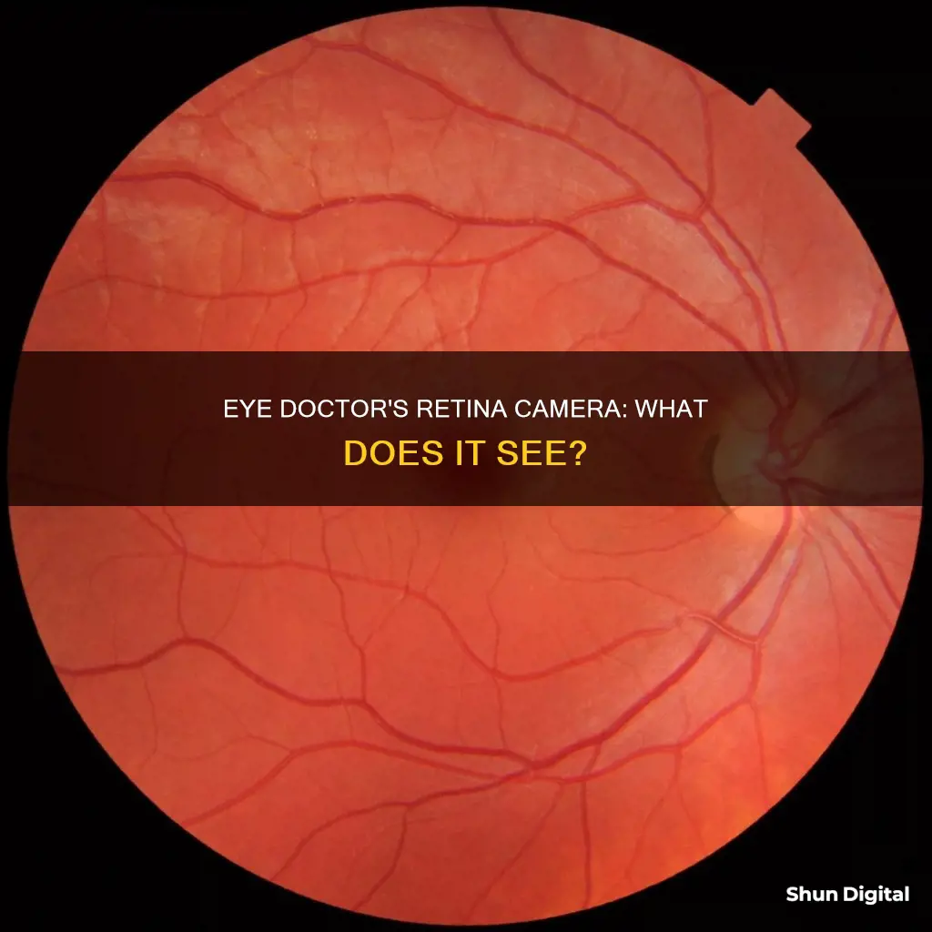
Retinal imaging is a diagnostic test that uses a high-resolution camera to take digital pictures of the back of your eye, including the retina, optic disc, and blood vessels. This technique allows eye doctors to detect specific conditions, such as diabetic retinopathy, glaucoma, and macular degeneration, and monitor changes in eye health over time. While retinal imaging is often recommended for patients with certain eye conditions or risk factors, it can also help identify serious health issues like diabetes, cardiovascular disease, and cancer. The procedure is quick, painless, and non-invasive, providing valuable insights for early treatment and disease prevention.
| Characteristics | Values |
|---|---|
| What is retinal imaging? | A diagnostic test that creates high-quality digital images of the inner, back surface of your eye. |
| What does it show? | Retina, optic disc, optic nerve, macula, blood vessels |
| What can it detect? | Diabetes-related macular edema, diabetes-related retinopathy, macular degeneration, retinal vein occlusion, age-related macular degeneration, diabetic eye disease, glaucoma, retinal toxicity |
| What are the benefits? | Early detection of vision issues, quick, painless, effective, can be compared with previous images to monitor eye health and detect subtle changes |
| What are the risks? | Fluorescein angiography may cause temporary skin discoloration, a slight yellow tint to urine, rarely an allergic reaction |
What You'll Learn

Retinal imaging can be used to detect diabetic retinopathy
Retinal imaging is a diagnostic test that uses advanced technology to capture high-quality, digital images of the back of your eye, including the retina, optic nerve, and other vital structures. This non-invasive procedure is an invaluable tool for eye doctors, as it allows them to detect and monitor various eye conditions, including diabetic retinopathy.
Diabetic retinopathy is a common complication of diabetes that can lead to vision loss if left untreated. The condition arises when diabetes damages the blood vessels in the retina, causing them to leak fluid or bleed. This, in turn, can lead to swelling in the macular region, known as macular edema, and the growth of abnormal blood vessels, known as neovascularization.
Retinal imaging plays a crucial role in the detection and management of diabetic retinopathy. Through retinal imaging, eye care specialists can identify signs of diabetic retinopathy that may not be apparent during a regular eye exam. This includes microaneurysms, which are small areas of swelling in the blood vessels, and macular edema, which is the buildup of fluid in the macula, the central part of the retina. By detecting these changes early on, doctors can intervene with appropriate treatments to prevent further damage and vision loss.
Additionally, retinal imaging is useful for monitoring the progression of diabetic retinopathy over time. Repeated imaging allows doctors to notice even subtle changes in the retina, helping them assess the effectiveness of treatments and adjust them as needed. This is especially important for people with diabetes, as the condition can cause progressive damage to the eyes if not properly controlled.
There are several methods of retinal imaging that can be used to detect and monitor diabetic retinopathy. These include color fundus photography, fluorescein angiography, optical coherence tomography (OCT), and widefield imaging. Each of these techniques offers unique advantages and provides valuable information to guide treatment decisions.
In summary, retinal imaging is a powerful tool that enables eye doctors to detect, diagnose, and manage diabetic retinopathy effectively. By utilizing this technology, doctors can identify signs of the condition early on, monitor its progression, and provide timely interventions to prevent vision loss in people with diabetes.
Android Smartwatches: Camera-Equipped or Not?
You may want to see also

Retinal imaging can help diagnose macular degeneration
Retinal imaging is a diagnostic test that uses advanced optical and camera technology to capture high-quality, detailed digital images of the inner, back surface of the eye, including the retina, optic nerve, and other critical structures. This non-invasive procedure is an invaluable tool for eye doctors, enabling them to detect and monitor various eye conditions, including macular degeneration.
Macular degeneration is an age-related eye disease that affects central vision, causing difficulty in seeing things directly in front of the affected individual. While peripheral vision remains intact, the condition can significantly impair the ability to perform everyday tasks. The condition primarily affects those over 50, with almost 20 million adults in the US currently affected. By 2040, it is predicted that this number will rise to 288 million people worldwide.
The condition has two forms: dry (atrophic) and wet (exudative). Dry macular degeneration, accounting for about 90% of cases, is characterised by the formation of tiny yellow protein deposits called drusen under the macula. This leads to a gradual thinning and deterioration of the macula, resulting in a slow loss of central vision over time. Wet macular degeneration, on the other hand, is caused by the development of abnormal blood vessels under the retina and macula. These abnormal vessels leak blood and fluid, forming a bulge in the macula, which can lead to a rapid and severe loss of central vision.
Retinal imaging plays a crucial role in diagnosing and monitoring both types of macular degeneration. Optical Coherence Tomography (OCT), a significant advancement in retinal imaging, uses infrared light reflections off the retina to generate cross-sectional images. These images help eye doctors visualise pockets of fluid in the retina in patients with wet macular degeneration, guiding the frequency of necessary injections. OCT images also reveal drusen deposits, retinal thinning, and atrophy, which are indicative of dry macular degeneration.
Another imaging technique, retinal fundus photography, provides colour images of the retina, helping to detect drusen, blood, lipids, and atrophy associated with macular degeneration. Autofluorescence imaging and scanning laser ophthalmoscopy further enhance the detection of atrophy and provide detailed views of individual retinal cells.
The availability of advanced retinal imaging techniques has revolutionised the diagnosis and treatment of macular degeneration. These technologies enable eye doctors to identify early signs of the condition, monitor its progression, and guide appropriate treatments to slow or prevent vision loss.
Toshiba Fire TV: Camera-Equipped or Not?
You may want to see also

Retinal imaging can identify retinal detachment
Retinal imaging is a diagnostic test that uses technology to create high-quality digital images of the inner, back surface of your eye. This includes the retina, optic nerve, and other important structures. This technology can be used to identify retinal detachment, a serious eye condition where the retina (a light-sensitive layer of tissue at the back of the eye) is pulled away from its normal position.
There are several methods of retinal imaging, including color fundus photos, optical coherence tomography (OCT), and fluorescein angiography. These methods can identify retinal detachment by capturing images of the retina and other structures at the back of the eye. For example, OCT can show cross-sectional views of the layers of the retina, revealing any detachment.
Retinal detachment is a medical emergency that can lead to permanent vision loss or blindness if left untreated. The symptoms of retinal detachment include a sudden increase in floaters (dark spots or squiggly lines in your vision), flashes of light, and a shadow or "curtain" affecting your field of vision. If you experience any of these symptoms, it is important to see an eye doctor or go to the emergency room immediately.
Retinal imaging is a valuable tool in diagnosing and managing retinal detachment and other eye conditions. It allows eye doctors to identify detachment and monitor the condition over time, helping to prevent vision loss.
Simplisafe Camera Setup: Viewing Your Camera Footage
You may want to see also

Retinal imaging can be used to detect glaucoma
Retinal imaging is a diagnostic test that uses technology to create high-quality digital images of the inner, back surface of your eye. This includes the retina, the optic nerve, and other important structures. It is a valuable tool for the early detection and diagnosis of various eye conditions, including glaucoma.
Glaucoma is a progressive eye disease that can cause irreversible blindness if left untreated. It is characterized by damage to the optic nerve, which is responsible for transmitting visual information from the eye to the brain. Early-stage glaucoma typically goes unnoticed by patients as it has no effect on vision. However, as the disease advances, it leads to a loss of peripheral vision that can progress to blindness.
Retinal imaging plays a crucial role in the detection and management of glaucoma. While glaucoma cannot be diagnosed solely through retinal imaging, it provides valuable insights that form the foundation for an accurate diagnosis. Retinal images can capture signs of glaucomatous damage, such as changes in the optic disc and cupping of the optic disc, which is the increase in cup size relative to the optic disc.
There are various retinal imaging technologies available, such as color fundus photos, optical coherence tomography (OCT), and scanning laser polarimetry (SLP). These technologies offer high-resolution images that enable eye specialists to identify subtle changes in the retina over time. By comparing retinal images taken at different intervals, eye specialists can monitor the progression of glaucoma and determine the effectiveness of treatments.
The benefits of retinal imaging in glaucoma detection are significant. It provides an objective, quantitative, accurate, and precise assessment of the ocular structure. This helps eye specialists make more informed decisions about the presence and progression of glaucoma, leading to earlier treatment interventions. Additionally, retinal imaging reduces the need for repeated perimetric testing and brings the level of clinical interpretation closer to that of an expert observer.
In conclusion, retinal imaging is an invaluable tool for the detection and management of glaucoma. It enables eye specialists to visualize and analyze the retina, optic nerve, and surrounding structures, aiding in the early detection and diagnosis of this potentially blinding disease. While retinal imaging has its limitations, such as an inability to detect retinal bleeding, it remains a powerful adjunct to comprehensive eye examinations.
Accessing Geeni Cameras on PC: A Step-by-Step Guide
You may want to see also

Retinal imaging can be used to detect cancer
Retinal imaging is a diagnostic test that uses digital photography to capture high-quality images of the inner, back surface of your eye, including the retina, optic disc, optic nerve, and blood vessels. This non-invasive procedure is an important tool for eye doctors to assess eye health and detect various eye conditions, such as diabetic retinopathy, macular degeneration, and glaucoma.
But retinal imaging has broader applications beyond eye care. It can also aid in the early detection of certain types of cancer and other health issues, including stroke, cardiovascular disease, and hypertension. The detailed images obtained during a retinal exam provide a valuable record of an individual's eye health at different life stages, enabling doctors to identify subtle changes over time.
During a retinal exam, patients are directed to gaze into a device, one eye at a time, and a brief flash of light indicates that the image has been captured. The process is comfortable, quick, and painless, and the resulting images are immediately available for review by both the doctor and patient.
While retinal imaging does not replace traditional pupil dilation during an eye exam, it can complement it by offering a more comprehensive view of the back of the eye. This combination of retinal imaging and pupil dilation may enhance the detection of issues that could threaten vision, providing valuable information for determining a treatment plan to support long-term eye health.
In summary, retinal imaging is a powerful tool that not only helps eye doctors assess eye health and detect eye conditions but also contributes to the early identification of certain cancers and other health problems. By capturing detailed images of the back of the eye, this non-invasive procedure enables doctors to make more informed decisions about their patients' eye care and overall well-being.
Unraveling the Mystery of Person, Woman, Man, Camera, TV
You may want to see also
Frequently asked questions
A retina camera is a device used by eye doctors to take high-quality digital images of the inner, back surface of your eye, including the retina, optic nerve, and other important structures.
Retina cameras allow eye doctors to detect and monitor various eye conditions, such as diabetic retinopathy, macular degeneration, and retinal vein occlusion. Early detection and monitoring can help prevent serious disease progression and vision loss.
Retina cameras use low-power lasers or light waves to capture digital images of the retina. The light passes through the pupil to the retina, creating images that are collected by a machine, forming a detailed picture of the retina.
Eye doctors can see the retina, optic disc, and blood vessels. They can detect specific eye or health conditions, such as diabetic retinopathy, glaucoma, macular degeneration, retinal detachment, and signs of high blood pressure.
The frequency of retinal imaging depends on your individual needs and will be recommended by your eye care provider. Generally, it is advised to have imaging done annually, especially for those at high risk for retinal diseases or over the age of 5.


