It is not possible to see germs with the naked eye, but scientists have developed several ways to detect them. One method is to use a microscope, which can be a compound microscope or a phase-contrast microscope. Another way is to use a smartphone camera with a microscope add-on and a bacteria-detecting chip. A team of researchers from Duke University has also developed a 3D virus cam that can detect tiny viral germs. Additionally, there is a trick that involves using clear tape, markers, and the flash on a cellphone camera to turn it into a germ-finding black light.
| Characteristics | Values |
|---|---|
| Microscope | Required to see germs |
| Microscope type | Compound or phase-contrast |
| Microscope magnification | 400x to 1000x or more |
| Microscope lens | 4x, 10x, 40x, or 100x |
| Smartphone camera | Can be used with a microscope lens attachment |
| Stains | Methylene blue, crystal violet, or safranin |
| Germ appearance | Tiny dots, rods, or spirals |
What You'll Learn

Using a microscope lens and a smartphone camera
The light source used is a compact blue laser diode, powered by 3 AAA batteries. The laser is positioned to illuminate the sample at a steep, oblique angle to prevent background light from interfering with the detection of the sample signal. The device also includes a specialised filter to block background noise in the images. The sample is positioned in a holder on a miniature mechanical stage that can be moved to adjust the depth and focus. The built-in lens of the smartphone camera, along with an external lens, is used to magnify the sample, and the magnified fluorescent images are recorded by the sensor chip in the phone.
This method was used to detect fluorescent beads as small as 100 nm and fluorescently labelled human cytomegaloviruses, ranging from 150 to 300 nm in diameter. The researchers confirmed the ability of the device to detect nanoscaled objects by viewing the same samples with conventional scanning electron microscopy and confocal microscopy.
Another method to turn your smartphone into a germ-finding tool is by using clear tape, markers, and the flash on your camera. This can turn your phone into a black light to expose germs.
Stop Neighbors Spying: Block Their Camera Views
You may want to see also

Stains to enhance visibility
Staining is a technique used to enhance contrast in samples, usually at the microscopic level. Stains and dyes are used in histology, cytology, and medical fields such as histopathology, hematology, and cytopathology. Stains may be used to define biological tissues, cell populations, or organelles within individual cells.
In biochemistry, staining involves adding a class-specific dye (DNA, proteins, lipids, or carbohydrates) to a substrate to qualify or quantify the presence of a specific compound. Staining and fluorescent tagging can serve similar purposes.
In vivo staining, also called vital staining or intravital staining, involves dyeing living tissues to reveal their form or position within a cell or tissue. In vitro staining involves colouring cells or structures that have been removed from their biological context.
There are two types of staining techniques: simple staining and differential staining. Simple staining uses only one type of stain on a slide at a time, whereas differential staining uses multiple stains per slide.
- Crystal violet stains both Gram-positive and Gram-negative organisms. Treatment with alcohol removes the crystal violet colour from Gram-negative organisms only.
- Safranin is used as a counterstain to colour Gram-negative organisms that have been decolorised by alcohol.
- Methylene blue is used to stain animal cells, such as human cheek cells, to make their nuclei more observable.
- Congo Red Capsule Stain is a modification of the nigrosin negative stain. The bacteria take up the congo red dye, and the background is then stained with acid fuchsin dye.
- Gram Stain is a differential stain used to divide specimens of bacteria into two groups: Gram-negative and Gram-positive. The primary stain is crystal violet, followed by a mordant (iodine), a decolorizing agent (Gram's decolorizer or ethanol), and a counterstain (safranin).
- Negative staining is a mild technique that does not destroy microorganisms and is therefore unsuitable for studying pathogens. It uses a black or dark dye, such as nigrosin or India ink, to stain the background instead of the organisms.
- Simple stains, such as methylene blue, can be used to determine the morphology of fusiform and spirochetes in oral infections.
It is important to note that staining is not limited to biological materials. It can also be used to study the structure of other materials, such as semi-crystalline polymers or block copolymers.
Additionally, there are products available, such as Glo Germ, that can be used as a visual tool to teach proper handwashing and infection control practices. These products use gel, oil, or UV lights to demonstrate handwashing, surface cleaning, and hygiene techniques.
Dressing for the Camera: What to Wear on TV
You may want to see also

Using a smartphone app
While there are no smartphone apps that allow you to see germs with your camera, there are a few ways in which your smartphone can be used to detect bacteria.
Smartphone Microscope Adapter
One way to use your smartphone to detect bacteria is by attaching a microscope lens adapter to your phone. This method was tested by researchers at the University of Massachusetts Amherst, led by food scientist Lili He. The process involves using a bacteria-detecting chip and a microscope lens adapter to test samples of water, juice, or mashed vegetable leaf for bacterial load. The chip, used with a light microscope for optical detection, relies on a "capture molecule" called 3-mercaptophenylboronic acid (3-MBPA) that attracts and binds to any bacteria. The smartphone app visually detects the bacteria in the samples containing the chip. This method can detect as few as 100 bacteria cells per 1 millilitre of solution.
Smartphone-Based Pathogen Diagnosis
Another method, developed by researchers at the University of California, Santa Barbara, uses a smartphone app and a lab kit to identify bacteria from patients. The app uses the phone's camera to measure a chemical reaction and determine a diagnosis in about an hour. This method was specifically designed for the diagnosis of urinary tract infections but can also be modified to detect other pathogens and diverse patient specimens (blood, urine, and feces). The app is free and can be downloaded from the Google Play Store.
DIY Cellphone Black Light
A simple do-it-yourself method to turn your cellphone camera into a germ-finding black light involves using clear tape, a few markers, and the flash on your cellphone camera. However, it is unclear how effective this method is in actually detecting germs.
See-Through Trailers: Chevy's Truck Camera System Explained
You may want to see also

The need for high magnification
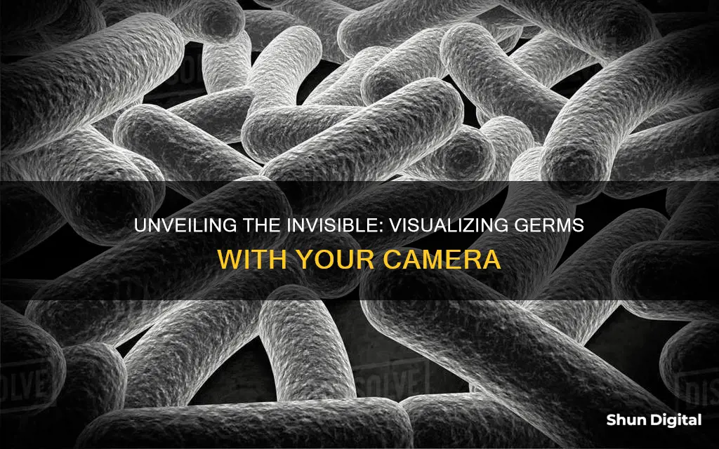
To see germs, you need to use a microscope as they are too small to be observed by the naked eye. Most bacteria are 0.2 micrometres in diameter and 2-8 micrometres in length, and they come in a variety of shapes, from spheres to rods and spirals. Single bacteria are transparent, so they are difficult to see even under a microscope.
To calculate the total magnification of a compound microscope, you need to know about two sets of lenses: the objective lens and the eyepiece lens. The objective lens is the one closest to the specimen slide and it produces an enlarged, inverted image of the specimen. The eyepiece lens magnifies this image further. The total magnification is calculated by multiplying the magnification of the objective lens by the magnification of the eyepiece.
Most educational-quality microscopes have a 10x eyepiece and three objectives of 4x, 10x, and 40x, providing magnification levels of 40x, 100x, and 400x. However, even at 100x, bacteria are still difficult to see in detail. To see anything interesting, you really need an electron microscope or a magnification of at least 1000x. At this level of magnification, you can fill the frame with a single bacterium and see basic shapes like rods or cocci.
Even at high magnifications, it takes a skilled person to differentiate bacteria from small dust and dirt particles that may be present on the slide. Some bacteria are found in bunches, which makes it difficult to see the individual cells. To make bacteria easier to see, you can use phase-contrast optics, which make the bacteria darker or lighter than the background, or you can stain the bacteria, although this may introduce artifacts.
Home Security: Watching Your Home from Another State
You may want to see also

The type of microscope used
Compound microscopes have an eyepiece lens (ocular) and multiple objective lenses mounted on a rotating nosepiece. The objective lenses offer different levels of magnification, typically ranging from 4x to 100x or more. To view bacteria, a higher magnification of 40x or 100x is required. The compound microscope provides excellent resolution, allowing for detailed observation of bacterial structures.
Another type of microscope that can be used to view bacteria is a phase-contrast microscope. This is a specialised type of compound microscope designed to enhance the contrast of transparent and colourless specimens, such as bacteria. It uses a phase plate in the objective lens to transform subtle changes in the refractive index of the bacteria into visible contrasts. This technique allows for the visualisation of bacteria without the need for staining, preserving their natural characteristics and behaviours.
In addition to these microscopes, there are also stereo or dissecting microscopes, which have a magnification of around 10-100x, and portable microscopes like pocket microscopes or the Foldscope, which can provide magnifications of up to 120x and 340x, respectively. However, compound microscopes are considered the best option for observing all types of microbes, including bacteria.
Troubleshooting Nightowl Cameras Not Showing on TV
You may want to see also
Frequently asked questions
You can't see germs with a regular camera, but you can turn your phone into a germ-finding black light using clear tape, markers, and your camera flash. Alternatively, you can use a microscope lens attachment with your smartphone camera to detect bacteria.
You can use a $30 microscope lens attachment with your smartphone to detect bacteria.
First, collect a sample of water, juice, or mashed vegetable leaf and place a chemical-based detection chip in with the sample. Then, place the chip on a microscope slide and secure it on the microscope stage. Lower the objective lens, adjust the focus, and increase the magnification. Observe the slide for bacteria, which can appear as tiny dots, rods, or spirals.
Bacteria can appear as tiny dots, rods, or spirals, depending on their shape. They are usually invisible to the naked eye and require a microscope to be seen.


