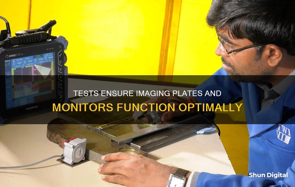
Tests performed on digital imaging plates and monitors are dependent on the type of imaging system being used. For example, in analogue radiography, tests include cassette tightness, inspections of the developing process, film storage conditions, film-screen contact tests, and anti-scatter grid tests. In computed radiography, tests include noise level, uniformity, anti-scatter grid, erasure thoroughness, high-contrast resolution, and defective pixel analysis.
Digital imaging plates and monitors are used in both computed radiography and digital radiography. In these systems, tests include noise level, uniformity, resolution, defective pixel analysis, and linearity and automatic response.
What You'll Learn
- Image quality parameters: detection efficiency, dynamic range, spatial sampling, contrast resolution, spatial resolution, noise, and quantitative detection efficiency
- Tests for imaging plates: noise, uniformity, anti-scatter grid, erasure thoroughness, resolution, low-contrast resolution, defective pixels analysis, and throughput
- Tests for monitors: resolution, colour accuracy, luminance, and uniformity
- Image processing: analysis, enhancement, and encoding
- Advantages of digital imaging: less radiation exposure, reduced time between exposure and image generation, ability to manipulate images, elimination of chemical processing, and ability to store patient records

Image quality parameters: detection efficiency, dynamic range, spatial sampling, contrast resolution, spatial resolution, noise, and quantitative detection efficiency
The quality of a digital image is determined by several parameters, including detection efficiency, dynamic range, spatial sampling, contrast resolution, spatial resolution, noise, and quantitative detection efficiency.
Detection Efficiency
The detective quantum efficiency (DQE) is a measure of the combined effects of the signal (related to image contrast) and noise performance of an imaging system. DQE is generally expressed as a function of spatial frequency. In medical radiography, DQE is used to describe how effectively an x-ray imaging system can produce an image with a high signal-to-noise ratio (SNR) relative to an ideal detector. A higher DQE means lower radiation exposure for patients. DQE is also important for CCDs, especially those used for low-level imaging in light and electron microscopy.
Dynamic Range
Dynamic range refers to the maximum variation in light intensity that a sensor can record. In digital imaging systems, dynamic range can be understood as the ratio between the luminance that produces white in the resulting image and the luminance that produces black. A camera's dynamic range helps us understand how well the system can reproduce scenes with high contrast, or large variations in brightness.
Spatial Sampling
The spatial resolution of a digital image is related to the spatial density of the image and the optical resolution of the microscope used to capture it. The number of pixels contained in a digital image and the distance between each pixel is known as the sampling interval, which is a function of the accuracy of the digitizing device. The Nyquist criterion states that the sampling interval must be equal to twice the highest specimen spatial frequency to accurately preserve the spatial resolution.
Contrast Resolution
Contrast resolution in radiology refers to the ability of an imaging modality to distinguish between differences in image intensity. The inherent contrast resolution of a digital image is given by the number of possible pixel values and is defined as the number of bits per pixel value.
Spatial Resolution
Spatial resolution in digital imaging refers to the number of pixels used to construct the image. Images with higher spatial resolution are composed of a greater number of pixels.
Noise
Noise refers to the random variation in the brightness or colour values of individual pixels in a digital image. It can be an issue when trying to pull significant detail out of extreme shadows or highlights by adjusting brightness and contrast values.
Quantitative Detection Efficiency
The DQE, as discussed above, is a quantitative measure of detection efficiency, taking into account the combined effects of signal and noise performance.
Straight Talk: Quick Monitor Alignment Check
You may want to see also

Tests for imaging plates: noise, uniformity, anti-scatter grid, erasure thoroughness, resolution, low-contrast resolution, defective pixels analysis, and throughput
Noise
Imaging plates can be susceptible to noise, which is the random variation in brightness or colour values of pixels. This can be an issue when attempting to extract significant details from extreme shadows in an image.
Uniformity
Uniformity testing in computed radiography involves evaluating the global uniformity index and detecting artifacts on the photostimulable phosphor screen. Uniformity of CR processing looks at each cassette’s imaging plate – fed independently in the CR reader. Imaging plates can be damaged over time due to artifacts, dust, dirt, debris and gears from the reader.
Anti-scatter grid
The anti-scatter grid enhances image quality in projection radiography by transmitting a majority of primary radiation and selectively rejecting scattered radiation. It is typically manufactured with lead strips oriented along one dimension, separated by a low attenuating interspace material.
Erasure Thoroughness
Erasure Thoroughness refers to the process of erasing any residual energy from imaging plates before they are returned to service. This is done by exposing the plates to intense white light to release any remaining energy that could affect future exposures.
Resolution
Resolution refers to the ability of imaging plates to distinguish between two separate points. This is determined by the pixel density of the digital data, which is in turn determined by the sampling frequency of the analog signal.
Low-contrast resolution
Low-contrast resolution refers to the ability of imaging plates to distinguish between shades of grey. This is determined by the pixel bit depth, which controls the number of grey shades that can be displayed.
Defective pixels analysis
Defective pixels in imaging plates can interfere with subtle features in medical images, reducing the quality of diagnosis. Each pixel in an LCD display has its own individual transistor that controls the transmittance of that pixel. Occasionally, these transistors malfunction, resulting in a defective pixel that always shows the same brightness.
Throughput
Throughput refers to the number of imaging plates that can be processed in a given amount of time. This can be improved by using multiwell plates, which are the gold standard for high-throughput screening and high content screening in cell culture.
Hyundai Accent: Blind Spot Monitoring Feature Explained
You may want to see also

Tests for monitors: resolution, colour accuracy, luminance, and uniformity
Tests for monitors include assessments of resolution, colour accuracy, luminance, and uniformity.
Resolution
Resolution is the ability of a monitor to display fine details. It is typically measured in pixels, the tiny dots that make up an image on a screen. The higher the resolution, the more pixels there are, and the sharper the image appears. Resolution can be measured using physical measurements, such as the number of pixels per inch (PPI) or dots per inch (DPI), or by using visual tests, such as viewing images with fine details or text at different resolutions.
Colour Accuracy
Colour accuracy refers to how well a monitor can reproduce colours accurately. This is important for tasks such as photo editing, where accurate colour representation is crucial. Colour accuracy can be measured using colourimeters or spectrophotometers, which analyse the colours displayed on the screen and compare them to a reference colour space, such as sRGB or Adobe RGB.
Luminance
Luminance refers to the brightness of a monitor. It is typically measured in candelas per square meter (cd/m^2) and can vary depending on the settings and the type of panel used in the monitor. Luminance can be measured using a luminance meter or a light meter, which measures the amount of light emitted by the screen.
Uniformity
Uniformity refers to how consistent the image quality is across the entire screen. This includes assessing factors such as brightness uniformity, colour uniformity, and viewing angle stability. Brightness uniformity can be measured using a luminance meter to check for variations in brightness across the screen. Colour uniformity can be checked by displaying a solid colour across the screen and looking for any variations or discolouration. Viewing angle stability can be assessed by viewing the screen from different angles to ensure that the image quality remains consistent.
Understanding the ASUS ACPI Monitor Application
You may want to see also

Image processing: analysis, enhancement, and encoding
Image processing involves performing operations on an image to enhance it or to extract valuable information from it. The two types of methods used for image processing are analog image processing and digital image processing.
Digital image processing is a subcategory of digital signal processing and has many advantages over analog image processing. It allows a much wider range of algorithms to be applied to the input data and can avoid problems such as noise and distortion.
There are three general phases that any type of data has to go through while undergoing digital processing:
- Enhancement and display
- Information extraction
- Encoding
The most common analysis operation is a histogram, which is a graphic representation of the number of pixels with a specific grey value. Brightness, contrast, and dynamic range data can be obtained from this analysis.
The most common enhancement operations are:
- Contrast manipulations
- Spatial filtering
- Subtraction
- Pseudo-colour
Image coding permits faster transmission of image data or better utilisation of a storage device. There are two basic types of encoding schemes: lossless algorithms, where no information is lost, and lossy algorithms, where information is lost.
Tommie's Ankle Monitor: A Question of Legal Intrigue
You may want to see also

Advantages of digital imaging: less radiation exposure, reduced time between exposure and image generation, ability to manipulate images, elimination of chemical processing, and ability to store patient records
Digital imaging is a technique used to record radiographic images without the use of film or processing chemistry. Instead, it uses an electronic sensor and a computerised imaging system to produce radiographic images almost instantly.
Advantages of Digital Imaging
Less Radiation Exposure
Digital imaging can reduce radiation exposure to the patient by 50-70% compared to traditional radiography.
Reduced Time Between Exposure and Image Generation
The processing speed of digital radiography allows technicians to produce an image for immediate review, increasing workflow efficiency.
Ability to Manipulate Images
The digital format allows for greater imaging versatility and overall image quality, increasing the accuracy of patient diagnosis and the quality of care. The image can be lightened or darkened, and annotations can be added.
Elimination of Chemical Processing
Digital imaging eliminates the need for chemical processors, processor maintenance, and the space required for dark rooms and cabinets of analog images. It also reduces environmental costs by removing the need for disposal of processing chemicals, silver salts in film emulsion, and lead foil sheets.
Ability to Store Patient Records
Digital imaging allows for the electronic storage of patient records.
Practical Tips for Buying Parts to Fix Split Monitors
You may want to see also
Frequently asked questions
Different imaging techniques used to pick up the signal of interest in digital sensors include charge-coupled devices (CCD), complementary metal-oxide semiconductors (CMOS), photostimulable phosphors plates (PSP) and tuned-aperture computed tomography (TACT).
Digital radiography sensors are divided into two types: storage phosphor plates (SPP) and silicon devices such as charge-coupled devices (CCD) or complementary metal oxide semiconductors (CMOS).
There are two types of digital sensor array designs: area and linear. Area arrays are used for intraoral radiography, while linear arrays are used in extraoral imaging.


