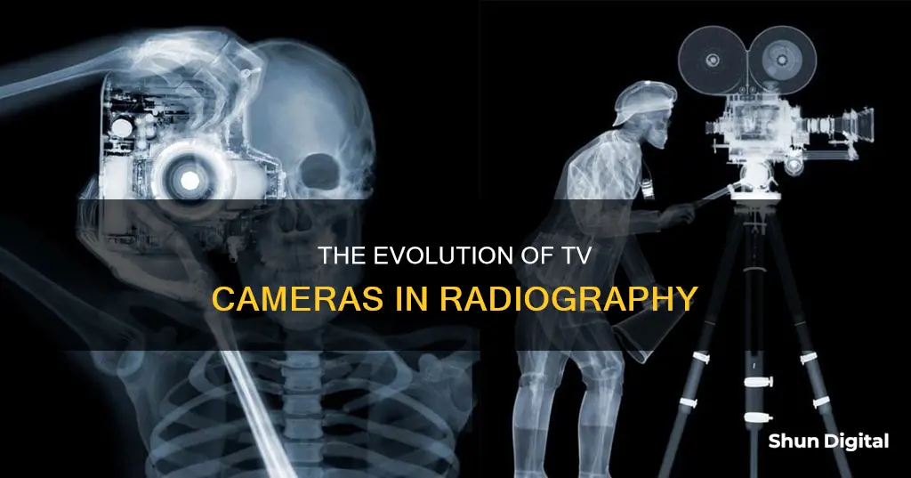
Radiography is a non-destructive testing method that uses radiation to create images of things that cannot be seen by the naked eye. In industrial radiography, both gamma rays and X-rays are used to inspect the integrity and structure of materials. Gamma rays are emitted from radioactive materials contained within the equipment, while X-rays are produced by applying a high voltage between the cathode and anode of an X-ray tube. These radiation types can travel through many substances, allowing inspectors to examine objects internally without altering them.
The pictures produced from radiographic testing are called radiographs, and most cameras used today record digital images. However, in the past, radiographs were captured using film.
| Characteristics | Values |
|---|---|
| Type of camera | Video camera, surveillance camera, analogue camera, digital camera |
| Functionality | Captures images and records videos |
| Storage | Video tape recorder, desktop computer, laptop computer, digital video recorder |
| Image Quality | Multi-megapixel, forensic quality, DVD quality |
| Resolution | 320,000 pixels, 640x480 pixels, 768x576 pixels, 720x480 pixels, 800x600 pixels, 1280x960 pixels |
| Frame Rate | 30 frames per second |
What You'll Learn

X-ray and gamma-ray radiation sources
X-rays and gamma rays are both forms of high-frequency, high-energy ionizing radiation. They are grouped together due to their shared properties and health effects. Both types of radiation can be naturally or artificially produced.
Natural Sources of X-Rays and Gamma Rays
X-rays and gamma rays can be produced by radon gas, radioactive elements in the Earth, and cosmic rays from outer space.
Artificial Sources of X-Rays and Gamma Rays
X-rays and gamma rays are created in nuclear power plants and used in medical imaging tests, cancer treatment, and food irradiation.
X-Rays
X-rays are emitted by electrons outside the nucleus of an atom. They can be produced naturally or by machines using electricity. X-ray machines are commonly used in medicine for medical imaging tests and cancer treatment.
Gamma Rays
Gamma rays are a form of penetrating electromagnetic radiation that arises from the radioactive decay of atomic nuclei. They are created by nuclear decay and have the shortest electromagnetic wavelength. Gamma rays are produced by nuclear reactors and high-energy physics experiments.
Industrial Gamma-Ray Sources
Iridium-192 and cobalt-60 are two common industrial gamma-ray sources used for radiography. These isotopes emit radiation at specific wavelengths and produce high energies that can penetrate thick materials with short exposure times.
Health Effects of Gamma Rays
Gamma rays are a health hazard and can cause DNA mutations, cancer, and tumours. They can also lead to radiation sickness, bone marrow damage, and internal organ damage.
Radiation Protection
Gamma rays require shielding with dense materials such as lead or concrete to reduce their intensity and protect from radiation exposure.
Unleashing Adobe Camera Raw: Editing Powerhouse for Photographers
You may want to see also

Detectors and their recordings
Detectors play a crucial role in radiography, capturing photons after they have passed through the specimen being examined. The choice of detector depends on the specific application and the type of radiation used, such as X-rays or gamma rays. Here are some commonly used detectors and the methods by which they record images:
- Silver Halide Film: This traditional method involves exposing photosensitive film to radiation. The film captures a latent image, which is then chemically developed to create a visible radiograph. This technique was commonly used before the advent of digital radiography.
- Phosphor Plate: Phosphor plates, also known as imaging plates or storage phosphor plates, are coated with photostimulable phosphors. When exposed to radiation, these plates store energy in the form of trapped electrons. To read the image, the plate is scanned with a laser beam, causing the stored energy to be released as light, which is then detected and converted into a digital image. This method offers reusability and higher sensitivity compared to traditional film.
- Flat Panel Detector: Flat panel detectors are digital devices that directly convert incoming radiation into electrical signals, producing high-quality images. These detectors consist of a scintillator layer that absorbs radiation and emits light, and an array of photodiodes that convert the light into electrical signals. Flat panel detectors provide superior image quality and faster acquisition times compared to other methods.
- CdTe Detector: Cadmium Telluride (CdTe) detectors are solid-state, direct-conversion detectors commonly used for X-ray and gamma-ray imaging. These detectors consist of a layer of CdTe crystals coupled with an array of electrodes. When radiation interacts with the CdTe layer, it generates electron-hole pairs, and the resulting electrical signals are captured and processed to form an image. CdTe detectors offer excellent spatial resolution and high detection efficiency.
- Digital Sensors: With the advancement of technology, digital sensors have largely replaced traditional film-based radiography. Digital cameras equipped with CCD (Charge-Coupled Device) or CMOS (Complementary Metal-Oxide-Semiconductor) sensors capture images by converting incoming radiation into electrical signals. These sensors offer superior image quality, higher dynamic range, and immediate access to images without the need for chemical processing.
It is worth noting that the choice of detector depends on factors such as the type of radiation used (X-rays or gamma rays), the energy level of the radiation, the required spatial resolution, and the specific application. Each detector has its advantages and limitations, and selecting the appropriate detector ensures accurate and reliable results in radiographic inspections.
Unlocking Macro Mode: How to Check Your Camera
You may want to see also

Radiation safety
Understanding Radiation Risks:
Radiation exposure, even in small amounts, carries potential risks for both patients and healthcare workers. X-rays and other forms of ionizing radiation can cause biological effects through deterministic and stochastic effects. Deterministic effects occur when specific exposure thresholds are exceeded, leading to radiation-induced conditions like thyroiditis, dermatitis, and hair loss. Stochastic effects, on the other hand, are probability-based and include the development of cancer years after radiation exposure.
Radiation Protection Principles:
The three fundamental principles of radiation protection are justification, optimization, and dose limitation. Justification involves weighing the benefits against the risks of radiation exposure for a particular procedure. Optimization aims to minimize radiation doses while still achieving the desired imaging results. Dose limitation entails adhering to established dose limits and the As Low as Reasonably Achievable (ALARA) principle to reduce radiation exposure as much as practicable.
To ensure radiation safety in radiography:
- Utilize radiation protection equipment: This includes personal protective equipment (PPE) such as lead aprons, thyroid shields, and leaded eyeglasses. Portable rolling shields and ceiling-suspended lead acrylic shields can also be employed to reduce radiation exposure for medical staff.
- Maintain distance: Increasing the distance between the X-ray source and the object being imaged helps minimize radiation exposure. The image intensifier or X-ray plate should be positioned as close to the patient as possible, with the X-ray tube placed farther away.
- Reduce exposure duration: Preplanning the required images, minimizing the use of magnification, and employing pulsed fluoroscopy instead of continuous fluoroscopy can help decrease exposure time.
- Educate hospital staff: Providing training on radiation safety practices and best practices can significantly enhance radiation safety in medical facilities.
- Monitor radiation exposure: Dosimeters should be worn by all staff who encounter ionizing radiation to measure their cumulative radiation exposure. This data is crucial for assessing and managing radiation safety.
- Ensure proper storage and testing of equipment: Lead aprons and other protective gear should be regularly inspected and stored properly to maintain their effectiveness.
- Comply with regulations and guidelines: Adhering to radiation safety guidelines and regulations set by competent authorities is essential to minimize risks and ensure the safe handling of radioactive materials.
Industrial radiography, which utilizes X-rays or gamma rays to inspect materials and components, poses unique radiation safety challenges. Here are some additional considerations for this field:
- Use appropriate shielding: The type of shielding material depends on the type of radiation being used. Sand, lead, steel, spent uranium, tungsten, and water are commonly used for shielding in industrial radiography.
- Work in pairs and use safety equipment: Industrial radiographers often work in pairs and are required to use specific safety equipment, including radiation survey meters, alarming dosimeters, gas-charged dosimeters, and film badges or thermoluminescent dosimeters.
- Clear the area of bystanders: Before conducting any testing, ensure that the nearby area is cleared of all personnel to prevent accidental exposure to radiation.
- Follow regulations and obtain permits: Radiographers may need permits, licenses, and specialized training to comply with local regulations and safety standards.
- Handle radioactive sources with care: The use of strong gamma sources in remote sites requires careful handling and secure storage to prevent "lost source" accidents, which can have severe consequences for public safety.
Benz-Gant Camera: Battery-Operated or Not?
You may want to see also

Radiographic testing
RT has several advantages over other NDE techniques. It is highly reproducible, can be used on a variety of materials, and the data gathered can be stored for later analysis. It is also effective and requires very little surface preparation. Additionally, many radiographic systems are portable, which allows for use in the field and at elevated positions.
There are two main types of RT: conventional radiography and digital radiographic testing. Conventional radiography uses a sensitive film that reacts to the emitted radiation to capture an image of the part being tested. This image can then be examined for evidence of damage or flaws. However, films can only be used once and they take a long time to process and interpret. On the other hand, digital radiography doesn't require film. Instead, it uses a digital detector to display radiographic images on a computer screen almost instantly. It allows for a much shorter exposure time and higher-quality images.
Commonly used digital radiography techniques in the oil, gas, and chemical processing industries include computed radiography, direct radiography, real-time radiography, computed tomography, and automated radiographic testing.
Smart Doorbell Camera: How Long Does the Battery Last?
You may want to see also

Industrial radiography equipment
Industrial radiography is a non-destructive testing method that uses ionising radiation to examine materials and components. It is used to identify and quantify flaws and deterioration in material properties that could cause engineering structures to fail. The process involves passing radiation through an object and capturing the resulting photons with a detector. This can be done in 2D or 3D, and in real-time or after image reconstruction.
Industrial radiography uses either X-rays or gamma rays. X-rays are produced by X-ray generators, which apply a high voltage between the cathode and anode of an X-ray tube. Gamma rays are generated by the natural radioactivity of sealed radionuclide sources. After passing through the object, the radiation is captured by a detector, such as a silver halide film, phosphor plate, flat-panel detector, or CdTe detector.
There are a variety of industrial radiography equipment and supplies, including:
- Darkroom supplies
- Digital plates
- Film illuminators
- Developing tanks
- Control assemblies
- Source tubes
- Field safety instruments
- Film digitizers
- Magnetic particle and liquid penetrant
- Welding inspection
- Casting parts inspection
- Food inspection
- Luggage control
- Sorting and recycling
- Aircraft maintenance
- Ballistics
- Turbine inspection
- Surface characterisation
- Coating thickness measurement
- Drug control
Unraveling Camera Battery Composition: What Powers Photography?
You may want to see also
Frequently asked questions
TV cameras in radiography are devices that use radiation to create images of things that cannot be seen by the naked eye.
The two primary types of TV cameras used in radiography are gamma-ray and X-ray cameras. Gamma-ray cameras are typically smaller and do not require electricity, whereas X-ray cameras are larger and can be turned on and off.
Gamma-ray cameras contain radioactive material within the equipment.
X-ray cameras are powered by electricity.


