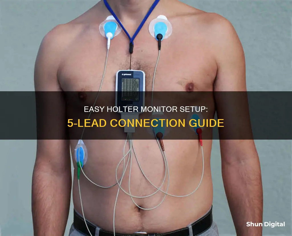
A Holter monitor is a type of portable electrocardiogram (ECG) that records the electrical activity of the heart over 24 hours or longer. It is often prescribed to patients experiencing irregular cardiac symptoms such as dizziness, fainting, and low blood pressure. The monitor is usually worn for 24 to 48 hours, during which the patient must keep a diary of their activities and symptoms. The device has wires that connect to electrodes placed on the chest and abdomen, and it must be kept dry at all times. The process of setting up a five-lead Holter monitor involves preparing the patient's skin, placing the electrodes, and connecting the wires to the monitoring device.
| Characteristics | Values |
|---|---|
| Number of leads | 5 |
| Purpose | Monitor electrical activity in the heart |
| Time worn | 24-48 hours |
| Placement | Chest and belly |
| Components | Adhesive electrodes, wires, monitor box |
| Diary | Required to log activities and symptoms |
| Charging | May require charging or battery change |
| Water contact | Must be kept dry |
What You'll Learn

Preparing the skin for electrode placement
Before placing the electrodes on the skin, the skin must be prepared to ensure the electrodes stay attached. First, identify the electrode sites and thoroughly shave all body hair from these areas. Next, rub the skin with an abrasive pad or lint-free gauze pad until the skin appears pink and dry. Then, clean and rub the skin with an alcohol prep pad until a reddish tinge appears. Let each site dry naturally and thoroughly.
After the skin is prepared, attach the lead wires by snapping the patient lead wires onto the electrodes. Peel the protective backing off the electrode and attach it to the patient, gently setting the gelled centres against the skin. Use care to avoid pressing gel out onto the adhesive. Smooth the electrode from the centre to the outer edge. If the electrode wrinkles as it is being applied, do not continue; replace the electrode with a new one.
Monitoring App Data Usage: Take Control of Your Mobile Data
You may want to see also

Positioning the electrodes
The correct placement of electrodes is crucial for accurate ECG interpretation. Inaccurate placement can distort the appearance of the ECG waveform, leading to potential misdiagnosis and mistreatment. The two major factors that determine the views of the ECG deflection on the monitor are the location of the electrodes on the body and the direction of the cardiac impulse in relation to the position of the electrode.
Firstly, assist the patient in lying down in the supine position and help them remove clothing that covers the chest. Then, identify the sternal angle or angle of Louis by locating the juncture of the clavicle and the sternum (suprasternal notch) and sliding your fingers down the center of the sternum to the obvious bony prominence, which is the sternal angle. The sternal angle identifies the second rib and provides a landmark for locating the fourth intercostal space (ICS) for accurate placement of electrodes.
For the five-lead system, follow these steps for electrode placement:
- Apply the Right Arm (RA) electrode close to the patient's right shoulder, below the clavicle.
- Apply the Left Arm (LA) electrode close to the patient's left shoulder, below the clavicle.
- Apply the Right Leg (RL) electrode just below the rib cage on the right side.
- Apply the Left Leg (LL) electrode just below the rib cage on the left side.
- Apply the chest lead electrode on the selected site: V1 at the fourth ICS right sternal border or V6 at the fifth ICS left midaxillary line. Only one precordial lead may be displayed, so the placement of this electrode identifies the lead used.
It is important to note that the electrodes should be placed tightly to prevent external influences from affecting the ECG. Press around the entire edges of the electrodes instead of directly on the gel pads, as pressing on the gel may cause leakage, which can interfere with transmission. Additionally, ensure that the patient's skin is cleaned and dried before placing the electrodes.
Tracking Techniques: Monitor Size Surveillance
You may want to see also

Connecting the electrodes to the monitor
Firstly, the electrodes should be connected to the lead wires before placing them on the patient's body. This is because placing the electrodes on the chest first and then attaching the wires may be uncomfortable for the patient. It could also lead to the formation of air bubbles in the electrode gel, which could interfere with the transmission of impulses.
Next, the patient's skin should be washed with soap and water, and dried thoroughly with gauze pads or a washcloth. This step is important because moist skin is not conducive to electrode adherence. Alcohol should not be used for skin preparation as it tends to dry out the skin.
After preparing the skin, the electrodes can be placed on the patient's chest and abdomen. The exact placement of the electrodes will depend on the type of system being used (three-lead, five-lead, or six-lead). For a five-lead system, the RA electrode is placed close to the right shoulder, below the clavicle; the LA electrode is placed close to the left shoulder, below the clavicle; the RL electrode is placed just below the rib cage on the right side; the LL electrode is placed just below the rib cage on the left side; and the chest lead electrode is placed on the selected site: V1 at the fourth intercostal space (ICS) right sternal border or V6 at the fifth ICS left midaxillary line.
Once the electrodes are in place, the lead wires should be plugged into the patient cable correctly and securely. The manufacturer's colour, letter, or symbol codes should be followed to ensure proper connection. For a five-lead system, the right arm wire plugs into the opening marked RA (usually white); the left arm wire plugs into the opening marked LA (usually black); the left leg wire plugs into the opening marked LL (usually red); the right leg wire plugs into the opening marked RL (usually green); and the chest wire plugs into the opening marked C or V (usually brown).
Finally, any tension on the lead wires and cables should be reduced to minimise interference or faulty recordings. For hardwire monitoring, a stress loop can be created by fastening the lead wire and patient cable to the patient's gown.
LCD Monitor Distortion: When and Why Does It Happen?
You may want to see also

Activating the monitor
There are various types and brands of Holter monitors, so instructions for activating the device may vary. Always follow the instructions provided by your doctor. The monitor will record continuously and turn off automatically when the prescribed period is over.
Make sure you see the activity lights flashing on the monitor itself so you know that it is working properly. The monitor will record every single heartbeat and can give information on the minimum, maximum, and average heart rate.
If your provider says it is okay, you can return to your normal activities once you have been hooked up to the monitor box and given instructions. This will let your provider find problems that may only happen with certain activities.
You may be told to keep a diary of your activities while wearing the monitor. Write down the date and time of your activities, especially if any symptoms such as dizziness, palpitations, chest pain, or other previously experienced symptoms occur.
Monitoring Oracle Temp Tablespace Usage: Tips and Tricks
You may want to see also

Returning the monitor
Before returning the monitor, you should remove it from your body. Dispose of the electrodes and place the device back into its original packaging or box. If you have any skin irritation or sticky residue from the electrodes, your doctor may have a solvent to help with this, or you can use water, alcohol, mineral oil, or lotion to remove the marks.
Along with the monitor, return any diary or log of your symptoms and activities during the monitoring period. This will help the cardiologist interpret the data from the monitor and make a more accurate diagnosis.
Finally, schedule a follow-up appointment with your cardiologist to discuss the results of the Holter monitor test. They will interpret the data and discuss any potential issues or treatment options.
LCD vs. LED Monitors: Which is Superior?
You may want to see also
Frequently asked questions
A Holter monitor is a type of portable electrocardiogram (ECG) that records the electrical activity of the heart over 24 hours or longer. It is used to evaluate symptoms that may be related to heart rhythm, identify irregular heartbeats, and assess the risk of future heart-related events.
Before attaching the electrodes, wash your skin with soap and water to ensure the adhesive pads stick properly. If you have thick chest hair, shave the areas where the electrodes will be attached to ensure a clear reading.
Place the adhesive electrodes on your body as directed by your doctor. Typically, this includes two electrodes on each side of your heart, one in front of your heart, and two pads on the bottom edge of each side of your rib cage. Secure the wires to the electrodes, and ensure the battery is charged before activating the device.


