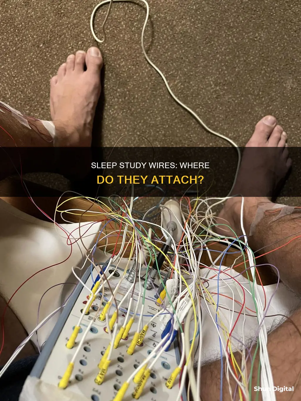
Sleep studies, or polysomnograms, are diagnostic tests that involve attaching sensors and electrodes to different parts of the body to monitor brain and body activity during sleep. The wires and sensors are attached to the scalp, face, chest, legs, fingers, and earlobes. These components monitor brain activity, eye movements, muscle activity, heart rhythm, body movements, nasal/oral airflow, respiratory effort, and oxygenation in the bloodstream. The data collected from these sensors helps physicians diagnose sleep disorders such as sleep apnea, restless leg syndrome, insomnia, and sleepwalking.
| Characteristics | Values |
|---|---|
| Brain activity | Electrodes are placed on the scalp |
| Eye movements | Electrodes are placed near the eyes |
| Muscle activity | Electrodes are placed on the face and a leg |
| Heart rhythm | Electrodes are placed on the chest |
| Body movements | Electrodes are placed on the chest and abdomen |
| Nasal/oral airflow | A nasal cannula is placed in the nose |
| Oxygen saturation | A clip is placed on the finger or earlobe |
What You'll Learn

Electrodes on the scalp
Electrodes are placed on the scalp to measure electrical brain activity. This is known as an electro-encephalogram (EEG). These electrodes are typically attached to your scalp close to the top (central), back (occipital) and frontal areas of your brain. The EEG is used to identify if you are awake, asleep, and the different stages of sleep you progress through.
EEG sensors have a sticky, electrically conductive gel coating that helps them stick to your head while they detect and record brain activity. Before applying the electrodes, the sleep technician will rub your scalp with an abrasive lotion and then scoop wax onto each electrode. This creates a tight seal and promotes conductance.
The placement of the electrodes on the scalp is very precise. The precise locations of the electrodes allow sleep technicians to study electrical activity from different parts of the brain. Each brain region shows characteristic wave patterns, which enable the technicians to identify what stage of sleep the participant is experiencing. They can even tell when the participant is dreaming!
The wires connecting the electrodes to a computer are usually gathered over the head with plenty of slack so that the participant can move around during sleep. After all the electrodes and wires are set up, the only thing left for the participant to do is go to sleep.
Surviving the Dating Game with an Ankle Monitor
You may want to see also

Sensors on the face
EMG sensors are placed on the skin, usually on the face and a leg, to track muscle movement. They monitor the electrical muscle activity produced by skeletal muscles. There is a lack of muscle tone during REM sleep, which is measured by the EMG channel. It is also used to test for medical abnormalities and sleep disorders such as Periodic Leg Movements of Sleep (PLMS).
EOG sensors are placed on the skin around the eyes to detect eye activity. Two sensors are placed around each eye, for a total of four sensors. They are used to identify sleep onset with slow rolling eye movements correlated with the EEG and REM sleep.
The sensors are attached to the body using medical tape and/or paste. They are not painful and are similar to the electrodes used in an electrocardiogram (EKG). However, they are for monitoring purposes only and do not activate any muscles.
Finding HP Pavilion P6 Series Monitor Specs
You may want to see also

Sticky pads on the chest
During a sleep study, a sleep technician will attach several wires and sensors to your body to monitor your sleep patterns. One of these sensors is a small, sticky pad that is attached to your chest via a wire. This electrode is used in electrocardiography (EKG) to monitor your heart rate and rhythm while you sleep.
The EKG sensor is coated with a sticky, electrically conductive gel that helps it adhere to your chest. This gel is skin-friendly and designed to keep the sensor in place throughout the night without causing any discomfort or irritation. The wires connecting the EKG sensor are long enough to allow you to move around comfortably in bed.
In addition to the EKG sensor, technicians may also attach other sensors and electrodes to your body, including those for electroencephalography (EEG), electromyography (EMG), and electro-oculography (EOG). These sensors work together to monitor your brain activity, muscle movement, eye movement, and other physiological parameters that provide valuable insights into your sleep patterns and quality.
Before the sleep study, it is important to follow the preparation instructions provided by your healthcare provider. This may include avoiding certain products or activities that can interfere with the sensors, such as creams, lotions, or caffeine. Inform your provider about any skin allergies you may have, as some adhesives used with the sensors may cause irritation or an allergic reaction.
Overall, the EKG sensor attached to your chest during a sleep study is an important tool for monitoring your heart function while you sleep, contributing to the comprehensive evaluation of your sleep patterns and quality.
Ankle Monitors: Secretly Recording Conversations?
You may want to see also

Elastic belts on the chest and abdomen
Elastic belts are often wrapped around the chest and abdomen to measure breathing during a sleep study. This is known as respiratory inductance plethysmography (RIP) belt technology. The belts contain a conductor that, when placed on a patient, forms a wire loop that creates inductance that is directly proportional to the absolute cross-sectional area of the body part it encircles.
The cross-sectional area is modulated with the respiratory movements and therefore also the inductance of the belt. By measuring the belt's inductance, a value is obtained that is modulated directly proportional to the respiratory movements. This is an inductance measurement of conductive belts that encircle both the thorax and abdomen of a patient.
Using two belts in a sleep study has considerable advantages for diagnostics. It gives the specialist a vital tool in estimating flow, which would not be possible with only one belt. Additionally, observing both ventilatory degrees of freedom gives the clinician a good picture of the patient's breathing during the study. As previously discussed, the patient can breathe through two different mechanisms: by "expanding the chest" and breathing "into the stomach". Having two points of reference for these mechanisms is critical during a sleep study.
The American Academy of Sleep Medicine (AASM) polysomnography guidelines recommend the use of RIP belts but not piezoelectrode (PE) belts for measuring chest and abdominal movements. However, some research suggests that PE belts perform similarly to RIP belts at distraction distances up to 10.0 centimetres.
Dismantling a Flat-Screen LCD Monitor: Step-by-Step Guide
You may want to see also

Pulse oximeter on the finger
A pulse oximeter is a small adhesive sensor that is attached to the tip of the index finger during a sleep study. It is used to measure the pulse and the level of oxygen in the blood. This is often used to check for respiratory problems and sleep apnea.
The pulse oximeter is a simple, non-invasive, and inexpensive method of monitoring blood oxygen levels. It is also known as an overnight oximetry test and can be done at home. It is a useful screening tool for sleep apnea and can be used to evaluate whether someone has a more common sleep disorder. It can also be used to monitor the response to continuous positive airway pressure (CPAP) treatment.
The pulse oximeter is a small plastic clip that is placed over the end of the finger. It is usually held in place with a piece of tape and is connected by a cable to a small box that records the data. Newer devices may adhere directly to the skin. The oximeter contains a red light that shines through the finger or surface of the skin, and a sensor that measures the pulse and oxygen content of the blood. The oxygen content is determined by the colour of the blood, which will vary depending on the amount of oxygen it contains. Highly oxygenated blood appears redder, while blood that is poor in oxygen is bluer. This changes the frequency of the light wavelength that is reflected back to the sensor.
The pulse oximetry data is recorded continuously throughout the night and will result in a graph. A medical provider will review the data to determine if there are abnormal drops in oxygen levels, known as desaturations, which may be indicative of sleep apnea.
In general, it is considered abnormal if oxygen levels fall below 88%, which indicates a condition called hypoxemia. Desaturations to less than 80% are considered severe and may require treatment such as CPAP or bilevel therapy.
The overnight pulse oximetry test is a commonly used screening test that is simple to perform and provides basic information about an individual's sleep.
Balanced Cables for Studio Monitors: Are They Necessary?
You may want to see also
Frequently asked questions
A technician will apply small sensors to your head, face, chest, and legs. The wires connecting the sensors to a computer are usually gathered over your head with plenty of slack so you can move around during sleep.
The wires are used to monitor brain activity, heart rate, eye movements, muscle activity, body movements, nasal/oral airflow, respiratory effort, and oxygenation.
The wires are attached using medical tape and/or paste. The sensors have a sticky, electrically conductive gel coating that helps them stick to the skin.


