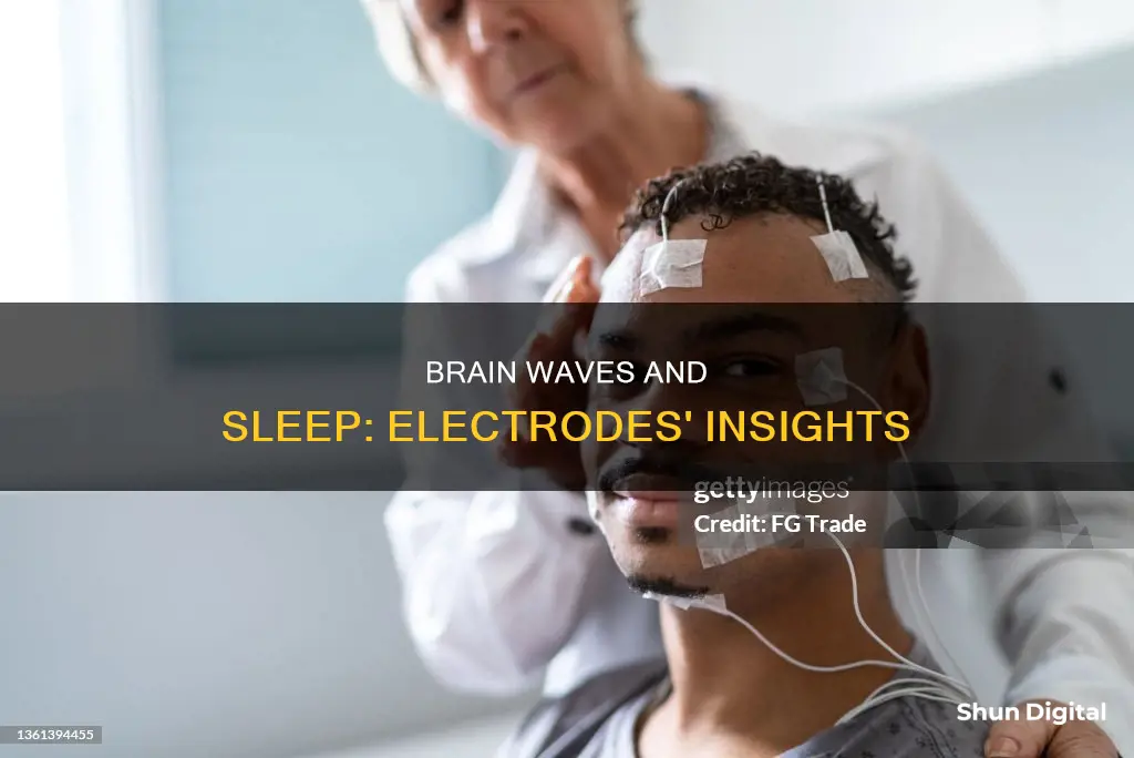
Electrodes monitor a variety of bodily functions during a sleep study, also known as a polysomnogram. These include brain activity, eye movements, muscle movement, heart rhythm, oxygen saturation, and more. The data collected from these electrodes helps sleep technicians and doctors diagnose and treat sleep disorders such as sleep apnea, restless leg syndrome, and insomnia. The process typically involves spending a night in a sleep lab or clinic, where the patient is hooked up to multiple electrodes and monitored while they sleep.
| Characteristics | Values |
|---|---|
| Brain Activity | Electrical Currents of the Brain |
| Eye Movement | Number of Eye Movements, Frequency or Speed |
| Limb Movement | Number and Intensity of Movements |
| Breathing Patterns | Number and Depth of Respirations |
| Heart Rhythm | Electrical Activity of the Heart |
| Oxygen Saturation | Percentage of Oxygen in the Blood |
| Acid/Base Balance of the Stomach | Amount of Acid Secreted During Sleep |
| Sleep Latency | Time Taken to Fall Asleep |
| Sleep Duration | Period of Time a Person Stays Asleep |
| Sleep Efficiency | Ratio of Total Time Asleep to Total Time in Bed |
What You'll Learn

Brain activity
Electrodes are small metal discs with wires attached that are placed on a patient's head and body to monitor brain waves, or brain activity, during a sleep study. This process is called an electroencephalogram (EEG). An EEG measures the electrical activity of the brain, producing a readout that can be "scored" into different stages of sleep.
During an EEG, a sleep technician will measure the dimensions of the patient's head and mark spots on the scalp and face where the electrodes will be attached. A mildly abrasive paste is applied to each spot to remove the oil from the skin so that the electrodes can adhere properly. The electrodes are then gently placed on the marked spots, and some of the wires may be taped in place.
For a typical EEG, six "exploring" electrodes and two "reference" electrodes are used. However, if a seizure disorder is suspected, more electrodes will be applied to document the appearance of seizure activity. The exploring electrodes are usually attached to the scalp near the frontal, central (top), and occipital (back) portions of the brain. These electrodes provide a readout of the brain activity that can be scored into different stages of sleep: N1, N2, and N3 – which together are referred to as non-rapid eye movement (NREM) sleep – and Stage R, which is rapid eye movement (REM) sleep, or wakefulness.
The EEG is one of the basic recordings done during a sleep study, which is also called a polysomnogram or polysomnography (PSG). A polysomnogram measures multiple parameters of sleep, including brain activity, eye movements, muscle activity, and heart rhythm. The test is used to help diagnose sleep disorders such as sleep apnea, restless leg syndrome, and narcolepsy.
Finding Epson Status Monitor 3: A Step-by-Step Guide
You may want to see also

Eye movement
Electrooculography (EOG) is a method of measuring eye movement during sleep. It is one of the sensors used in a sleep study, alongside others like the electrocardiogram (EKG) and the electromyography (EMG).
The EOG sensor is typically placed on the outer edges of each eye and is used to measure electrical eye movement activity. It is used to help identify sleep onset, with slow rolling eye movements correlated with the EEG (electroencephalography) and REM sleep. The standard sleep scoring manual recommends that an electrooculogram utilises at least two channels. One electrode is placed 1 cm superior and lateral to the outer canthus of one eye, with the input connected to an amplifier. Another electrode is placed 1 cm inferior and lateral to the outer canthus of the other eye, forming input 1 to a second amplifier. This placement scheme detects conjugate horizontal and vertical eye movements. For example, when the eyes look to the right, the cornea of the right eye approaches electrode A, and electrode A becomes positive relative to the inactive ear. According to the polarity convention, amplifier 1 registers a downward deflection.
Several varieties of eye movements are recorded during routine PSG (polysomnography). While the patient is awake, saccadic eye movements, as well as eye blinks, are noted. Eye blinks produce the same EOG pattern as vertical eye movement. One of the first signs of drowsiness is the cessation of any eye movements. As the patient gets sleepier, slow eye movements are seen. These usually have a frequency of less than 0.5 Hz, are most consistently recorded in the horizontal axis, gradually increase in amplitude as background alpha activity drops out, and usually disappear in stage 2 sleep.
During REM sleep, rapid eye movements (REMs) occur. Movements along the horizontal axis are the most common, though oblique and vertical movements occur as well. REMs typically occur in bursts and may be preceded on the EEG by characteristic “sawtooth” waves. There is no widely accepted definition of REMs that would serve to distinguish them from slow eye movements.
G-Sync Monitors: Performance Enhancers or Detractors?
You may want to see also

Heart rate
Electrodes are used to monitor heart rate during a sleep study, also known as nocturnal polysomnography (NPSG). Electrodes are attached to the body to monitor heart rhythm and rate. This is done using an electrocardiogram (EKG) or an electrocardiogram (ECG) measurement system. The ECG system uses conductive textiles on a bed for long-term healthcare monitoring. The EKG sensor uses potential electrical waveforms to measure heart rhythm and rate.
The heart rate is monitored to gain a better understanding of the quality and stages of sleep. There are four sleep stages that a person typically goes through, and each is important individually. The first stage is the transition from being awake to falling asleep, characterised by slow eye-rolling movements. The second stage is the beginning of consolidated sleep, where larger brain waves are displayed and there is a lack of eye movement. The third stage is a deep sleep where it is hard to arouse the sleeper, and they will display slow and large delta waves on the PSG. The fourth stage is REM sleep, where the eyes jerk around in different directions, the limbs become paralysed temporarily, and the heart rate increases.
The heart rate is also monitored to help diagnose sleep disorders, including sleep apnea, restless legs syndrome (RLS), and other sleep-related respiratory disorders. Sleep apnea is a total obstruction of breathing that lasts for more than 10 seconds. The severity of sleep apnea is determined by the Apnea Hypopnea Index (AHI), which measures the number of sleep apnea and hypopnea (partial obstruction) episodes during sleep.
Is Your Monitor G-Sync? Here's How to Tell
You may want to see also

Limb movement
During a sleep study, electrodes play a crucial role in monitoring various physiological parameters to assess sleep quality and identify any underlying sleep disorders. One of the key parameters they help evaluate is limb movement.
Electrodes and Limb Movement Monitoring
To accurately capture these movements, electrodes are strategically placed on the patient's limbs. These electrodes may be placed on the legs and, depending on the specific evaluation, may also be placed on the arms. The placement is determined by the suspected disorder and the patient's symptoms. The electrodes employed for this purpose are typically surface electrodes, similar to those used in standard electroencephalography (EEG) or electromyography (EMG) procedures.
Measuring Muscle Activity
The electrodes used for limb movement monitoring are designed to record electrical signals from the muscles, allowing for the detection of even subtle movements. This is crucial for understanding the frequency, intensity, and patterns of limb movements during sleep. For instance, in the case of PLMD, electrodes help identify periodic, abrupt, and repetitive limb movements that typically occur at intervals of 20–40 seconds during sleep.
Analyzing Limb Movements
The data gathered by the electrodes is meticulously analyzed to differentiate between normal and abnormal limb movements. This analysis considers factors such as the number of movements per hour and the amplitude or intensity of the movements. Additionally, the impact of these movements on sleep architecture is assessed. By correlating the frequency and timing of limb movements with the various stages of sleep, sleep specialists can make accurate diagnoses and formulate effective treatment strategies.
In summary, electrodes are indispensable for monitoring limb movements during sleep studies, providing valuable insights into potential sleep disorders. This information is pivotal for tailoring treatment plans aimed at improving sleep quality and alleviating symptoms associated with abnormal limb movements during sleep.
Blind Spot Monitoring: A Necessary Safety Feature?
You may want to see also

Sleep duration
Sleep studies, also known as polysomnography (PSG) or polysomnogram, involve recording various physiological parameters during sleep. The test is typically performed overnight in a sleep lab or clinic, and the patient is monitored by a trained technologist or technician.
During a sleep study, electrodes are attached to the patient's head and body to monitor different aspects of sleep. One of the key parameters measured is sleep duration, which refers to the period of time a person stays asleep. This is an important metric as it provides insights into the patient's sleep habits and sleep quality.
By measuring sleep duration, sleep specialists can determine if the patient is getting an adequate amount of sleep. The ideal sleep duration for adults is typically considered to be around 7-9 hours per night. However, the optimal sleep duration may vary slightly between individuals.
In addition to sleep duration, other parameters measured during a sleep study include sleep latency (how long it takes to fall asleep), sleep efficiency (the ratio of time asleep to time in bed), eye movement, brain activity, limb movement, breathing patterns, heart rhythm, and oxygen saturation.
All the data collected during a sleep study is then analysed by a sleep specialist, who interprets the results and discusses them with the patient. This information helps in diagnosing sleep disorders and developing appropriate treatment plans to improve sleep quality and duration.
Best Places to Buy Omron 3 Series BP Monitor
You may want to see also


